

J1: Super-Resolution Microscopy - DNA-PAINT
For more than a century, the diffraction limit set a hard boundary to what light microscopes could resolve: Structures closer together than 200 nm simply blur into one spot...
Super-resolution microscopy circumvents that barrier, and the underlying idea is surprisingly simple!
In this lab course you will experience it first hand by building and operating your own super-resolution microscope in a single day.
Supervising Tutors:
Thomas Kellerer (kellerer@biochem.mpg.de)
Martin Benoit (martin.benoit@physik.uni-muenchen.de)
Location:
Jungmann Lab
Max Planck Institute of Biochemistry
Am Klopferspitz 18, 82152 Martinsried
Further information:
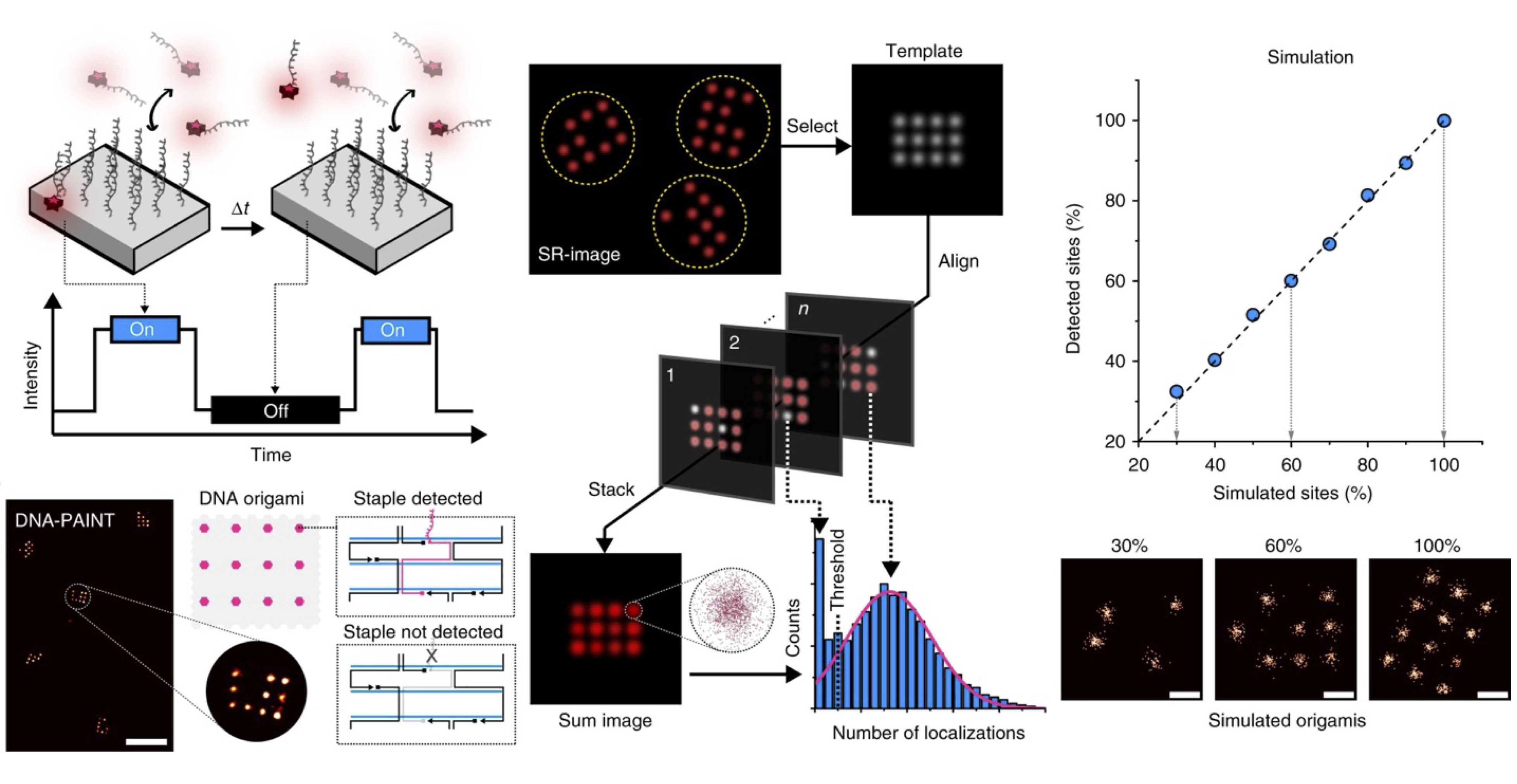
The course is built around DNA-PAINT, a method in which short fluorescently labeled DNA strands transiently bind to complementary strands on the sample. Each binding event produces a brief blink, and from many thousands of such blinks the position of every single molecule can be pinpointed with a precision far below the diffraction limit. Our samples are DNA origami nanostructures, self-assembled objects that carry binding sites in a defined pattern only a few tens of nanometers apart. Under a conventional fluorescence microscope they appear as featureless spots. With DNA-PAINT, the nanometer-sized patterns become visible.
The heart of the course is the microscope itself. You will work with a liteTIRF setup, a compact total internal reflection fluorescence microscope built from a small number of optical components. We will go through the optical path step by step, from the laser and the excitation geometry to the objective, the emission filters and the camera, and discuss why each part is needed and what happens when it is misaligned. The goal is to show how quickly and with how little equipment a super-resolution microscope can be assembled, and to generate a real feeling for the optics (compared to a typical microscope as a black box).
Once the setup is running, you will acquire DNA-PAINT data of DNA origamis and reconstruct super-resolved images from the raw image sequence. In the second part of the day we explore what actually determines the achievable resolution. You will vary parameters such as laser power, exposure time, imager concentration and acquisition length on the real microscope and compare the results with simulations, so that you can see directly which factors help, which ones hurt, and why.
The course is aimed at Bachelor students in physics, biophysics, chemistry or related subjects. No prior experience with microscopy or DNA nanotechnology is required. A basic understanding of geometrical optics and fluorescence is helpful.
Further reading:
Lelek, M., Gyparaki, M. T., Beliu, G., Schueder, F., Griffié, J., Manley, S., Jungmann, R., Sauer, M., Lakadamyali, M. and Zimmer, C., Single-molecule localization microscopy, Nature Reviews Methods Primers 1, 39 (2021).
https://www.nature.com/articles/s43586-021-00038-x
Auer, A., Schlichthärle, Th., Wöhrstein, J. B., Schueder, F., Strauss, M. T., Grabmayr, H. and Jungmann, R., Nanometer-scale multiplexed super-resolution imaging with an economic 3D-DNA-PAINT microscope, ChemPhysChem 19, 3024 (2018).
https://chemistry-europe.onlinelibrary.wiley.com/doi/10.1002/cphc.201800630
Schnitzbauer, J., Strauss, M. T., Schlichthärle, Th., Schueder, F. and Jungmann, R., Super-resolution microscopy with DNA-PAINT, Nature Protocols 12, 1198 (2017)
https://www.nature.com/articles/nprot.2017.024